
Shear wave elastography Simplify liver disease assessment
The value of elastography
Philips shear wave elastography simplifies liver assessment, making obtaining liver stiffness measurements fast and easy. This non-invasive, reproducible, and easily performed method of assessing liver tissue stiffness may help reduce, or even avoid, the need for conventional liver biopsies1. Research suggests that instead of a costly and painful biopsy procedure, an easy ultrasound exam could become routine for assessing liver disease status.
Elastography in liver assessment:
ElastPQ (P-SWE) has proven valuable in staging liver disease, reducing the need for costly and potentially painful biopsies in some patients. ElastQ (2D-SWE) advances shear wave elastography, providing a technique that is not only noninvasive and easy to use, but also offers clinicians additional confidence in the reliability of measurements, even for the technically difficult patient (TDP), such as a patient with a high BMI.
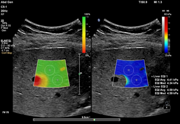
ElastQ imaging
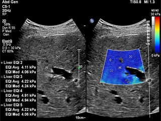
ElastQ imaging is a real-time, large Region of Interest (ROI), color-coded quantitative assessment of tissue stiffness. It is the only solution to offer both PureWave technology and high frame rate real-time shear wave imaging across abdominal applications. Clinicians can easily assess liver tissue stiffness using real-time feedback and make quantitative measurements with multiple sample points even retrospectively on DICOM stored images. Unique confidence map, used with the stiffness map, improve confidence of shear wave measurements. Both maps can be displayed side-by-side, which reduces workflow steps and allows for simultaneous map correlation during acquisition and measurement phases.
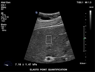
ElastPQ
ElastPQ is an easy-to-use method of obtaining tissue stiffness values of the liver on a predefined ROI. Using real-time imaging as a guide, the ROI is placed over the area of interest and tissue stiffness data such as AVG, MEAN, and IQR are obtained and displayed in seconds. Multiple samples can be recorded and liver tissue report generated from the results.
Shear wave elastography
Simplify liver disease assessment
The value of elastography
Philips shear wave elastography simplifies liver assessment, making obtaining liver stiffness measurements fast and easy. This non-invasive, reproducible, and easily performed method of assessing liver tissue stiffness may help reduce, or even avoid, the need for conventional liver biopsies1. Research suggests that instead of a costly and painful biopsy procedure, an easy ultrasound exam could become routine for assessing liver disease status. Philips shear wave elastography simplifies liver assessment, making obtaining liver stiffness measurements fast and easy. This non-invasive, reproducible, and easily performed method of assessing liver tissue stiffness may help reduce, or even avoid, the need for conventional liver biopsies. Research suggests that instead of a costly and painful biopsy procedure, an easy ultrasound exam could become routine for assessing liver disease status. Philips shear wave elastography simplifies liver assessment, making obtaining liver stiffness measurements fast and easy. This non-invasive, reproducible, and easily performed method of assessing liver tissue stiffness may help reduce, or even avoid, the need for conventional liver biopsies. Research suggests that instead of a costly and painful biopsy procedure, an easy ultrasound exam could become routine for assessing liver disease status. Philips shear wave elastography simplifies liver assessment, making obtaining liver stiffness measurements fast and easy. This non-invasive, reproducible, and easily performed method of assessing liver tissue stiffness may help reduce, or even avoid, the need for conventional liver biopsies. Research suggests that instead of a costly and painful biopsy procedure, an easy ultrasound exam could become routine for assessing liver disease status.

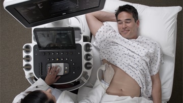
Noninvasive liver assessment made easy
The liver stiffness measurement is captured in seconds through a simple noninvasive scan

Easily assess and monitor patients
Shear wave elastography simplifies liver disease assessment
Simplify liver assessment with noninvasive tools Obtaining liver stiffness measurements with Philips shear wave elastography is surprisingly easy and fast even on difficult-to-image patients. It’s noninvasive, making it a quick, simple step for sonographers and virtually painless for patients.
What is it?
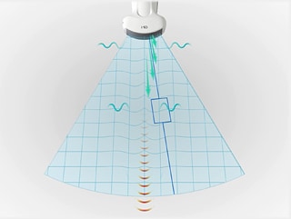
Philips elastography generates shear waves inside the liver by using acoustic force from a focused ultrasound beam. The system monitors shear wave propagation and measures its velocity, then displays it in a format that is easy to interpret.
Noninvasive liver fibrosis assessment
ElastPQ ultrasound shear wave elastography Richard G. Barr, MD, PhD, FACR, Diagnostic Radiology,
Hitchcock Imaging, Youngstown, OH
Noninvasive liver assessment made easy
The liver stiffness measurement is captured in seconds through a simple noninvasive scan
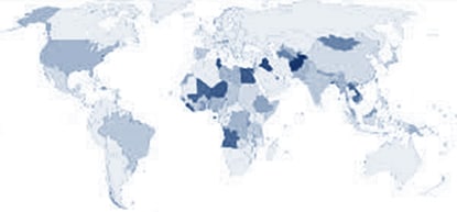
Chronic Hepatitis C Worldwide

1 Ferraioli G, et al. Point shear wave elastography method for assessing liver stiffness. World J Gastroenterology. 2014 April 28;20(16):4787-4796.
Easily assess and monitor patients
Shear wave elastography is supported on the EPIQ 7, EPIQ 5, and Affiniti 70 ultrasound systems.
1 Ferraioli G, et al. Point shear wave elastography method for assessing liver stiffness. World J Gastroenterology. 2014 April 28;20(16):4787-4796.