
Liver ultrasound
The ultimate solution for liver assessment
It’s no secret that liver disease is a growing health concern.
Globally, liver disease accounts for around 2 million deaths per year, with around 1 million of those due to cirrhosis and 1 million due to viral hepatitis and hepatocellular carcinoma. Unhealthy lifestyle choices like obesity, alcohol and drug use are the root causes of the disease and in most cases, liver disease can be prevented1.
For decades, liver biopsy has been the conventional method for disease assessment, and more recently MR Proton Density Fat Fraction (MR-PDFF) has been added to measure liver fat. This method, however, presents several patient barriers including the pain of biopsy, access to MR imaging, high cost and long wait times.
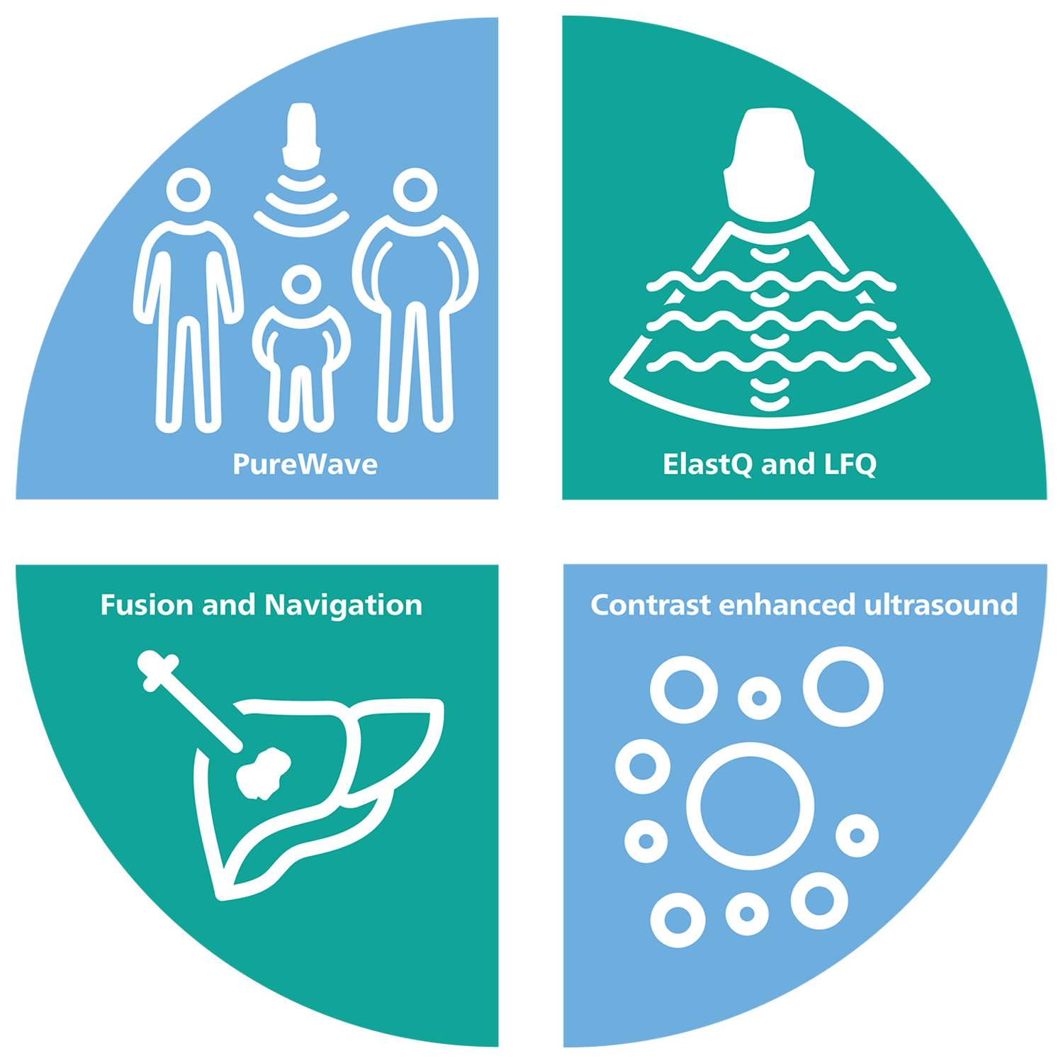
This is where Philips comes in. Thanks to innovations to Philips EPIQ & Affiniti ultrasound systems there is now a comprehensive solution to support the assessment, treatment and monitoring of liver disease, with four key features working together in one effective tool.
Quantitative parameters for assessment of steatosis
with Philips Liver Fat Quantification (LFQ) tools.
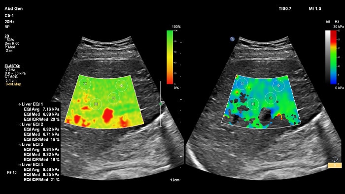
Easy, fast and robust acquisition
with ElastQ imaging.*
29% to 85% reduced pain and fatigue
from scanning with the C5-1 transducer compared to conventional transducers.
*Side-by-side view of unique imaging confidence and stiffness maps helps reduce workflow steps and allows for simultaneous map correlation during acquisition and measurement phases.
The widespread use of this technology in a general population could be helpful in screening for advanced chronic liver disease, especially considering that a complete study can be done in under three minutes using a non-invasive method for chronic liver disease.”
Richard G. Barr, MD, PhD
President, Radiology Consultant, Inc. (Youngstown, Ohio) Medical Director, Southwoods Imaging
Easy quantifiable tools to assess liver health

ElastQ imaging
ElastQ provides a 2D shear wave (2D-SWE) real-time assessment of tissue stiffness that is noninvasive, reproducible, and easy to perform. ElastQ also includes the ability to make retrospective measurements on stored images and includes a confidence map for adequate shear wave propagation display.
ElastPQ imaging
ElastPQ is a point shear wave (pSWE) method of obtaining tissue stiffness values of the liver. Using real-time imaging as a guide, the ROI is placed over the area of interest and tissue stiffness data is acquired in seconds. Multiple samples can be recorded and generated into a liver report.
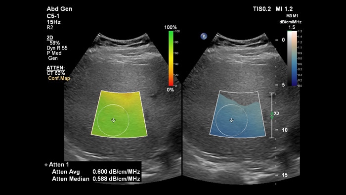
Attenuation imaging
Attenuation Imaging can measure the amount of fat present in the liver by calculating the attenuation coefficient of sound abnsorbed by the liver parenchyma. This method provides quantitative attenuation parameters that can help assist physicians in the management of patients with hepatic steatosis.
HRI imaging
HRI imaging technology compares the relative brightness of the liver parenchyma to the kidney and provide fast, quantifiable liver steatosis information.
Attenuation imaging
ElastQ imaging
Contrast enhanced liver ultrasound

Ultrasound contrast agents can transform the role of ultrasound, allowing clinicians to study the enhancement patterns of liver lesions in real time. With Philips ultrasound, contrast enhanced ultrasound is seamlessly integrated into the standard workflow.
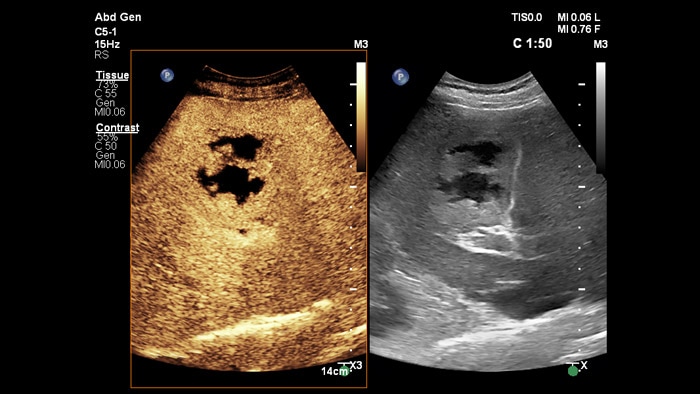
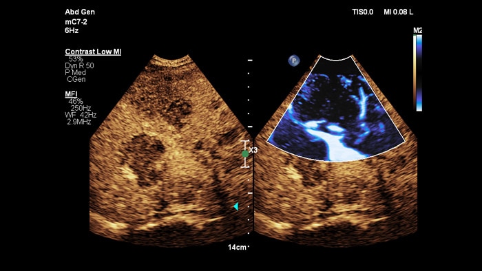
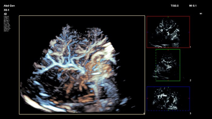
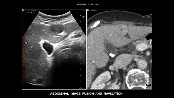
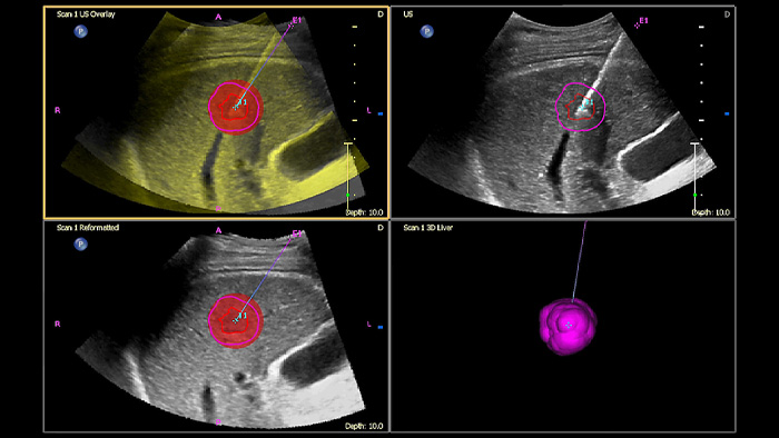
Image fusion and navigation

Make confident decisions even in challenging diagnostic cases with Philips image fusion and needle navigation capabilities. Streamlined workflow allows clinicians to achieve fast and effective fusion of CT/MR/PET with live ultrasound while needle navigation aids in guiding biopsy of small and difficult-to-access lesions2.
Using Tumor contour, users can plan and target lesions with a semi-automated tool that helps outline a 3D contour around a structure of interest, potentially improving procedure guidance with challenging cases where the lesion may not be easily visualized.
Transducers created to image all body habitus

People come in many different shapes and sizes, with PureWave crystal technology you can improve penetration in difficult to scan body types, all while maintaining excellent detail resolution, Doppler sensitivity and contrast enhanced ultrasound (CEUS) performance.
PureWave crystal technology is the biggest breakthrough in piezoelectric transducer material in 40 years and is 85% more efficient than conventional piezoelectric materials, resulting in exceptional performance for all patient body types.
Resources
Related products
-
EPIQ Elite
Philips EPIQ Elite ultrasound features an exceptional level of clinical performance, workflow, and advanced intelligence to meet the challenges of today’s most demanding practices. The EPIQ Elite platform brings ultimate solutions to ultrasound, with clinically tailored tools designed to elevate diagnostic confidence to new levels.
795098 -
Affiniti 70
The Affiniti 70 ultrasound system offers a powerful combination of performance and workflow for quick, confident diagnosis.
795210 -
Affiniti 50
Choosing a new ultrasound system is all about balance. You need accurate diagnostic information quickly, a simplified yet intuitive user interface, and easy access to critical features, along with an ergonomic design and the latest technology.
795208


References
1 Asrani et al, Burden of liver disease in the world. Journal ofhepatology, Vol 70, Issue 1, 2019.)
2 Kim E, Patel RS, Fischman AM, Nowakowski S, Lookstein RA. CT-Guided Liver Biopsy With Electromagnetic Tracking: Results From a Single-Center Prospective Randomized Controlled Trial. AJR 2014; 203:W715–W723)
Ultrasound portfolio extensions
Let Philips help you leverage the full potential of ultrasound over a wide range of clinical care areas with a comprehensive portfolio of performance-driven products, services and knowledge.