MR Qflow
Integrate Qflow as part of MR Cardiac suite2
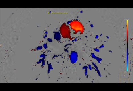
2Qflow still available also outside of CMR to accommodate non-cardiac use
Allow easy compare of flow results to cardiac function in ONE suite The application is designed to support visualisation and quantification of blood flow dynamics by reviewing MR Q-Flow data. The tooling creates 2D color flow overlay maps on anatomical references, e.g. to be used to calculate stroke volumes. The package includes automatic vessel contour detection for large vessels to quickly analyse vessel flow. Background correction allows for offset correction required for q-flow data of certain MR vendors.
MR Cardiac Quantitative Mapping
Map calculation for other acquisitions
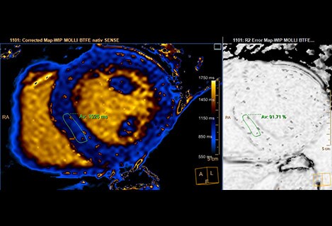
The application is designed to analyse quantitative maps provided by the scanner as well as calculated quantitative maps within the application for T1, T2 and T2* data. A toolbox is provided to manually improve the quantitative map calculation by manual and automatic motion correction tools. –Molli –shMolli –SASHA –T2prep
MR Cardiac Whole Heart
IntelliSpace Portal 10 – Task guided workflow
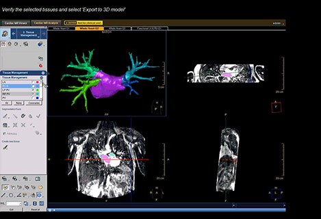
The application allows for 3D visualisation of any 3D or MRA scanned data. The application allows for automated segmentation of the heart in 3D data to provide a high quality 3D rendering of the heart. Furthermore it allows MRA data to be reviewed in Volume Rendering or MIP mode to segment relevant parts of the scanned data. With the tool the individual segmented tissues like left-ventricle, right-ventricle, coronaries etc. can be manually edited and selectively viewed and exported in a batch or saved as STL/VTK model to serve as 3D print or used in procedure planning. Features included: Volume Rendering, 3D heart segmentation algorithm, tissue list, (seed based) masking & segmentation and edit tools.
Enhancements in MR Cardiac
Easy comprehensive cardiac review for MR exams
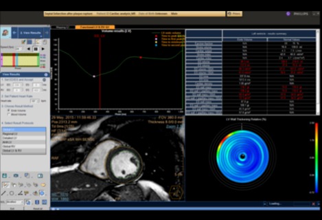
MR Cardiac enables quick visualisation with viewing protocols, of a single, multiple or all available cardiac series, including synchronisation of cardiac phases. The fast initial review allows clinicians to quickly view the general results of cardiac heart exams and decide on the first analysis that is needed. Visual scoring can be done using an AHA bull’s eye plot. Benefits: Allow multiple manual segmentation tools in LV ‒Freehand ‒Spline ‒Drop Seed Allow multiple manual segmentation tools in RV ‒Freehand ‒Spline
CT Cardiac Viewer
Quick cardiac visualisation
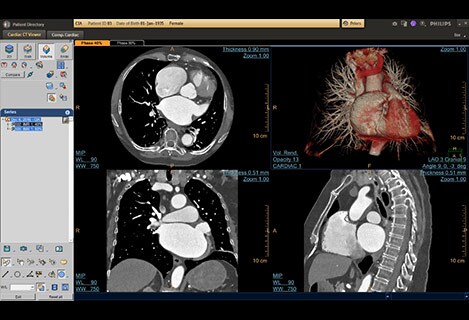
Rib cage removal for cardiac CT scans – enables a 3D anatomical volume rendering image of the heart and the large blood vessels connected to it, after removing the rib-cage structures automatically, for different types of clinical questions, and scanning protocols. Assisting in visualisation of complex anatomy, and for results sharing (e.g. with surgeons)