Intuitive, reproducible, AI-enabled echocardiography
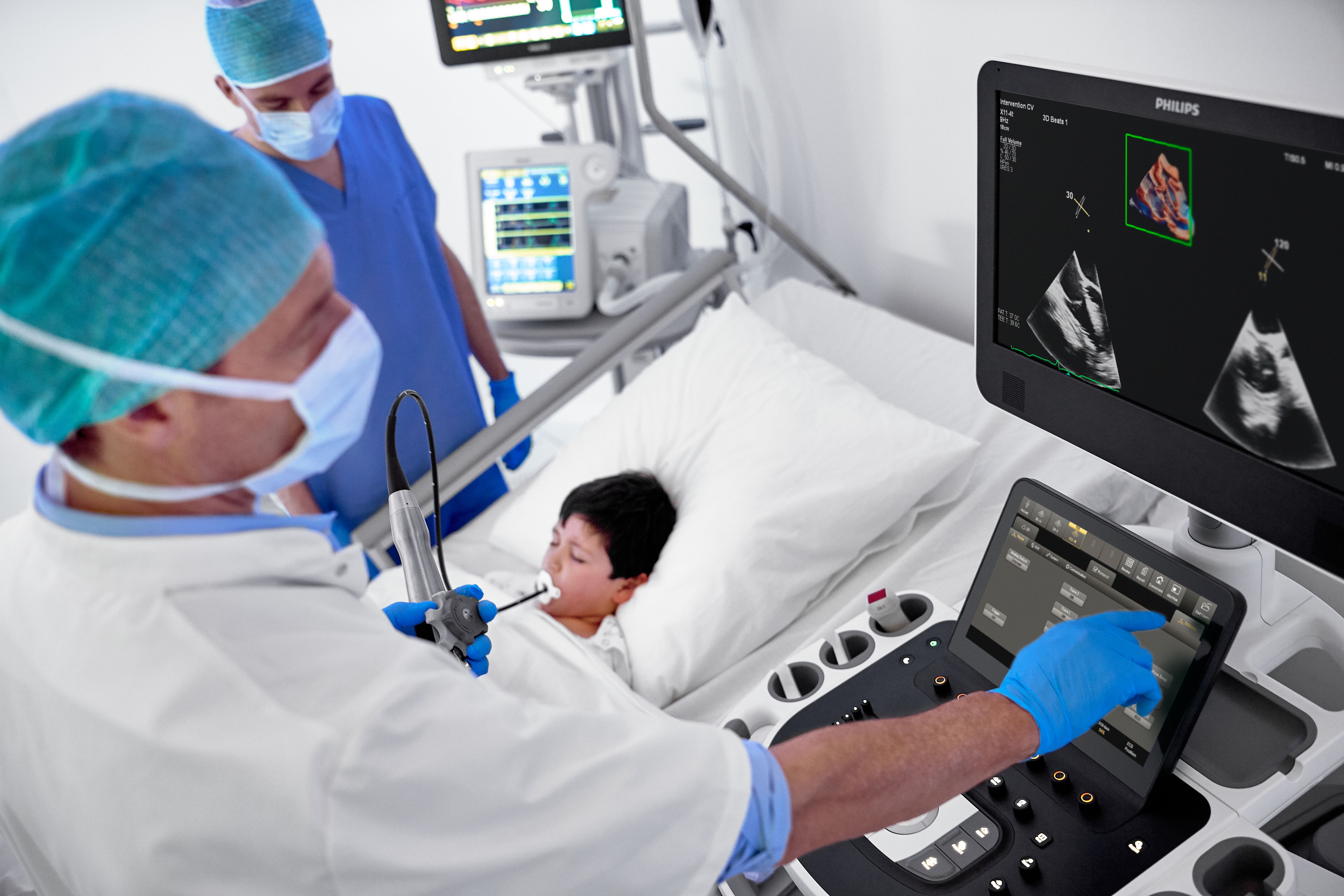
Powered by AI-based capabilities, our industry-leading CV ultrasound platform delivers advanced tools for structural heart disease, including 3D automated tricuspid valve and color flow quantification tools. Efficient automation and quantification tools enable you to diagnose heart failure and coronary artery disease quickly and confidently. Tiny transducers, high frame rates and sharp imaging support confidence in pediatric echo exams. Unified on- and off-cart workflow supports lab setup and consistent UI and workflow across systems.

White paper: Dynamic quantification of mitral regurgitation


