
Studying connectivity between brain regions
FieldStrength MRI magazine
Research - June 2017
The need for large amount of diffusion directions and high b-values
“From a research perspective the bottom line is that we want to use diffusion MRI to study connectivity between brain regions,” says Dr. Watts. “Previous diffusion methods, using a diffusion tensor imaging (DTI) model with a limited number of directions and typical b-values of 1000 s/mm2, are not able to support these kinds of quantitative connectome studies.”
Richard Watts, PhD, is Co-Director of the University of Vermont MRI Center for Biomedical Imaging, and an Associate Professor in the Department of Radiology. His academic interests include T1-weighted imaging in multiple sclerosis, Alzheimer’s disease, brain tumors, multimodal imaging of mild traumatic brain injury (mTBI), therapeutic hypothermia, and imaging connectomes using high b-value diffusion imaging.
"A greater number of directions and range of b-values opens up the ability to apply more sophisticated and biologically realistic models”
“Overcoming these limitations requires us to use high b-values to give us a couple of advantages. One is that we suppress extracellular water, so the signal has a much more specific interpretation, representing the highly anisotropic intra-axonal water content. Another advantage is that we get much higher angular resolution at higher b-values so we can determine a more accurate fiber orientation distribution within a voxel. The higher angular resolution requires us to sample more directions so that we can be sure to capture signals from directions where the fibers are close to perpendicular to the diffusion encoding gradient. And in more complex geometry with multiple fiber populations, we can better separate those individual fiber populations.”
Higher resolution with more sophisticated models
“There is a close analogy with digital camera technology; using high b-values is like having a higher quality lens, it produces a sharper image. But, to make the most of that sharpness, you need a higher resolution sensor, or, in the case of MRI, to measure more directions”.
“While a minimum of 6 directions, and a single b-value was sufficient for fitting the diffusion tensor model, measuring a much greater number of directions and a range of b-values opens up the ability to apply more sophisticated and biologically realistic models.” Reference 1. ABCD Study*: https://addictionresearch.nih.gov/abcd-study * Philips is not sponsoring this study
According to Dr. Watts, the institute is currently acquiring data for a multicenter study focusing on adolescent brain cognitive development (ABCD), using diffusion and functional MRI to examine the development of brain regions associated with reward and risk-taking behavior in children and adolescents.1
“We now employ a fairly sophisticated diffusion sequence with a total of 104 acquisitions at a range of b-values. We are now able to run this exam with more directions and at a higher spatial resolution than our previous protocol that we used in a study focusing on Alzheimer's, while maintaining the same scan time of about 10 minutes.”
Comparison of fiber tractography methods
Fiber tractography of the corticospinal tract with seed region of the posterior limb of the internal capsule. Different processing based on the same data.
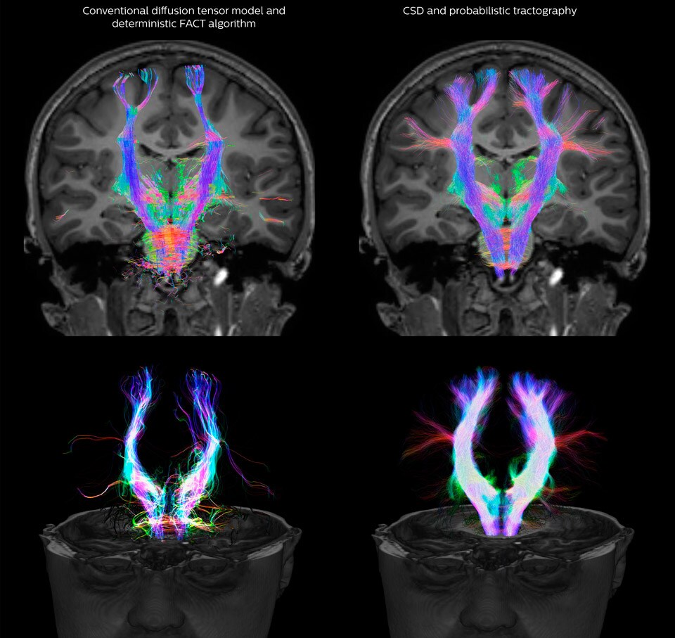
All images were created from the same acquisition in a child using Ingenia 3.0T CX and 32-channel dS Head coil. Diffusion data was acquired at b-values 0, 500, 1000, 2000, 3000. The use of high b-values (3000 s/mm2) effectively suppresses extra-axonal water signal and provides high angular resolution.
Legend of acronyms
CSD: constrained spherical deconvolution
DEC TDI: directionally encoded color track-density imaging
DTI: diffusion tensor imaging
DWI: diffusion-weighted imaging
FOD: fiber orientation density
Data processing was performed using open source software. Fiber tracking was performed using the MRtrix package (J-D Tournier, Brain Research Institute, Melbourne, Australia, https://github.com/MRtrix3/mrtrix3), Tournier et al. 2012. DEC TDI based on F Calamante et al 2010.
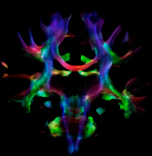
Fiber orientation density
CSD of multishell DWI results in the white matter FOD at each voxel. Unlike the conventional diffusion tensor model, this approach enables accurate modeling of multiple fiber populations within a single voxel.
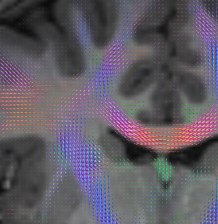
Crossings of the corpus callosum, corticospinal tracts, and the superior longitudinal fasciculus are shown.
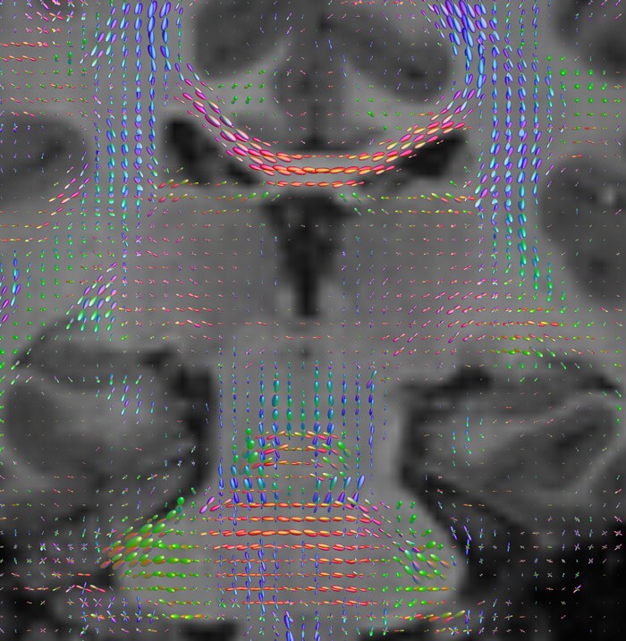
Crossing fibers of the corpus callosum bordered by the superior longitudinal fasciculus as well as within in the pons.
Fiber tracking based on CSD analysis of multishell DWI data and probabilistic tractography.
Fiber tracking from the left and right hippocampi to the fornix.
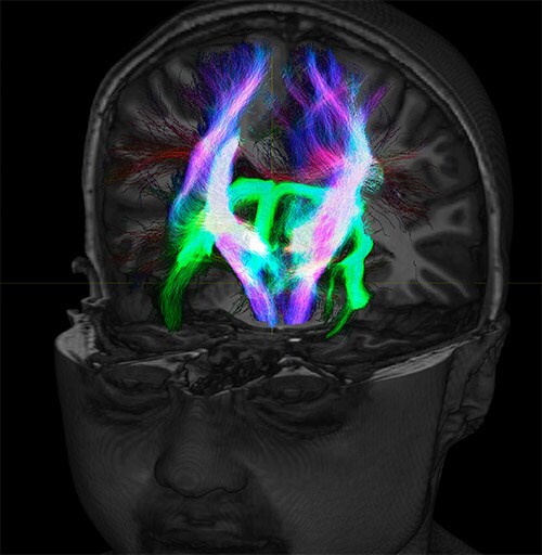
Fiber tracking from the left and right hippocampi to the fornix (green), and the corticospinal track based on a seed region of the posterior limb of the internal capsule.
DEC TDI
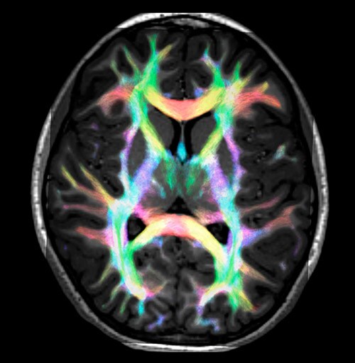
Super-resolution directionally encoded color track-density imaging overlaid on T1-weighted structural MRI.
Super-resolution directionally encoded color track-density imaging
Results from case studies are not predictive of results in other cases. Results in other cases may vary.

More from FieldStrength