- Get step-by-step guidance to simplify CBCT acquisition
-
Get step-by-step guidance to simplify CBCT acquisition
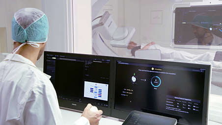
To help all clinical users [1] achieve superb 3D images, regardless of their level of experience, SmartCT Soft Tissue provides step-by-step guidance and visual aids during acquisition. This includes room set-up, isocentering the system, as well as suggesting a suitable contrast injection and X-ray acquisition protocol. - Interact with your CBCT image at table side
-
Interact with your CBCT image at table side
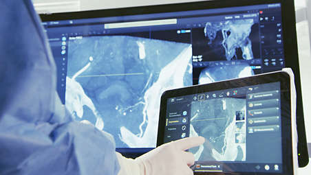
Once acquired, SmartCT Soft Tissue automatically displays the CBCT image on the touch screen module and the FlexVision within seconds for direct review at table side. - Access advanced 3D measurements at table side
-
Access advanced 3D measurements at table side
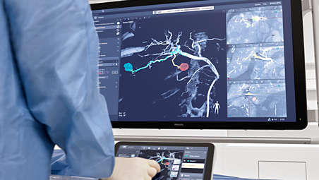
To study the type and extent of disease or anatomy of interest with great detail, you can render volumes, segment lesions and vessels, perform measurements and mark vessel paths to assess size and location of pathology or anatomy of interest directly on the TSM with the simplicity of a tablet gestures. - CBCT Open
-
CBCT Open
SmartCT Soft Tissue offers the possibility to acquire a CBCT using open trajectory with start and stop positions of +55° to -185° respectively. This protocol opens the arc to the left side of the patient allowing for a wider translation of the angiographic table towards this direction; thereby shifting the isocenter of the C-arm to the right lateral side of the patient. This enables visualizing off-centered regions of interest (such as the periphery of the liver) in a single sweep. [2] The Open arc trajectory also offers more comfort to the larger patients. - CBCT Dual in liver imaging
-
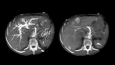
CBCT Dual in liver imaging
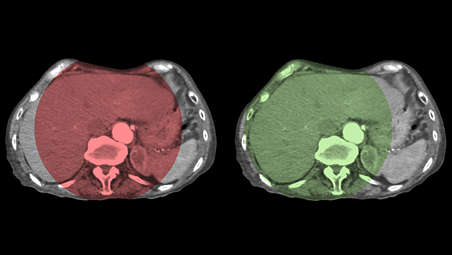
Dual Phase CBCT acquisition is an advanced capability of SmartCT Soft Tissue, which allows two CBCT scans to be made automatically on the Azurion system with a user defined interval and a single contrast injection. High resolution, high contrast images are reconstructed within seconds to support fast decisions during procedures. It is commonly used for TACE where the first phase serves as an arterial phase and the second (delayed) phase shows the contrast uptake in the lesions. [3,4] - CBCT Dual in brain imaging
-
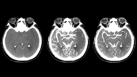
CBCT Dual in brain imaging
Dual phase CBCT is an increasingly used application for stroke diagnosis. A non-contrast CBCT aids detection of early ischemic changes. With CBCT Dual an early phase image helps to identify the proximal occlusion while the late phase supports the detection of collaterals, here again using a single contrast injection. [5]
Get step-by-step guidance to simplify CBCT acquisition
Get step-by-step guidance to simplify CBCT acquisition
Get step-by-step guidance to simplify CBCT acquisition
Interact with your CBCT image at table side
Interact with your CBCT image at table side
Interact with your CBCT image at table side
Access advanced 3D measurements at table side
Access advanced 3D measurements at table side
Access advanced 3D measurements at table side
CBCT Open
CBCT Open
CBCT Open
CBCT Dual in liver imaging
CBCT Dual in liver imaging
CBCT Dual in liver imaging
CBCT Dual in brain imaging
CBCT Dual in brain imaging
CBCT Dual in brain imaging
- Get step-by-step guidance to simplify CBCT acquisition
- Interact with your CBCT image at table side
- Access advanced 3D measurements at table side
- CBCT Open
- Get step-by-step guidance to simplify CBCT acquisition
-
Get step-by-step guidance to simplify CBCT acquisition
To help all clinical users [1] achieve superb 3D images, regardless of their level of experience, SmartCT Soft Tissue provides step-by-step guidance and visual aids during acquisition. This includes room set-up, isocentering the system, as well as suggesting a suitable contrast injection and X-ray acquisition protocol. - Interact with your CBCT image at table side
-
Interact with your CBCT image at table side
Once acquired, SmartCT Soft Tissue automatically displays the CBCT image on the touch screen module and the FlexVision within seconds for direct review at table side. - Access advanced 3D measurements at table side
-
Access advanced 3D measurements at table side
To study the type and extent of disease or anatomy of interest with great detail, you can render volumes, segment lesions and vessels, perform measurements and mark vessel paths to assess size and location of pathology or anatomy of interest directly on the TSM with the simplicity of a tablet gestures. - CBCT Open
-
CBCT Open
SmartCT Soft Tissue offers the possibility to acquire a CBCT using open trajectory with start and stop positions of +55° to -185° respectively. This protocol opens the arc to the left side of the patient allowing for a wider translation of the angiographic table towards this direction; thereby shifting the isocenter of the C-arm to the right lateral side of the patient. This enables visualizing off-centered regions of interest (such as the periphery of the liver) in a single sweep. [2] The Open arc trajectory also offers more comfort to the larger patients. - CBCT Dual in liver imaging
-
CBCT Dual in liver imaging
Dual Phase CBCT acquisition is an advanced capability of SmartCT Soft Tissue, which allows two CBCT scans to be made automatically on the Azurion system with a user defined interval and a single contrast injection. High resolution, high contrast images are reconstructed within seconds to support fast decisions during procedures. It is commonly used for TACE where the first phase serves as an arterial phase and the second (delayed) phase shows the contrast uptake in the lesions. [3,4] - CBCT Dual in brain imaging
-
CBCT Dual in brain imaging
Dual phase CBCT is an increasingly used application for stroke diagnosis. A non-contrast CBCT aids detection of early ischemic changes. With CBCT Dual an early phase image helps to identify the proximal occlusion while the late phase supports the detection of collaterals, here again using a single contrast injection. [5]
Get step-by-step guidance to simplify CBCT acquisition
Get step-by-step guidance to simplify CBCT acquisition
Get step-by-step guidance to simplify CBCT acquisition
Interact with your CBCT image at table side
Interact with your CBCT image at table side
Interact with your CBCT image at table side
Access advanced 3D measurements at table side
Access advanced 3D measurements at table side
Access advanced 3D measurements at table side
CBCT Open
CBCT Open
CBCT Open
CBCT Dual in liver imaging
CBCT Dual in liver imaging
CBCT Dual in liver imaging
CBCT Dual in brain imaging
CBCT Dual in brain imaging
CBCT Dual in brain imaging
Application areas

Lesion detection
DualPhase acquisition and DualView allow visualization of arterial and post-arterial contrast enhancement in liver imaging. With MRI-like lesion1,2 detection, XperCT Dual allows you to predict tumor response in TACE and SIRT procedures.

Neuro visualizations
XperCT Dual provides superb image quality when imaging areas where a coil or stent is present. Plus, its improved grey scale and more homogeneous images enhance visualization of soft tissue and small bleedings for post-procedural checks.

Visualization of endoleaks
The enhanced image quality of XperCT Dual improves the visualization of pathologies, such as endoleaks. It also assists in the treatment of large patients by providing improved image quality.
Related products
Alternative products
-
Azurion 7 M20
- Image Guided Therapy System Monoplane Ceiling/Floor Mounted with a 20" flat detector
- Enhance visibility for diverse vascular, oncology and cardiac procedures with great image quality
- Control all relevant applications via the central touch screen module at table side
View product
-
Azurion 5 M20
- Image Guided Therapy System Monoplane Ceiling/Floor Mounted with a 20” flat detector
- Covers a wide range of cardiac and vascular procedures to offer flexibility for multi-purpose use
- Control all relevant applications via the central touch screen module at table side
- Parallel working enables you to get more done, leading to high throughput and fast exam turnover
View product
-
SmartCT Vaso
- Step-by-step acquisition technique that can offer guidance to simplify 3D imaging
- Allows direct image inspection with advanced 3D visualization at table side
- Peri-procedure check of positioning of the flow-diverter stents
View product


-
Azurion 7 M20
Experience outstanding interventional cardiac and vascular performance on the Azurion 7 Series with 20'' flat detector. This industry leading image-guided therapy solution supports you in delivering outstanding patient care and increasing your operational efficiency by uniting clinical excellence with workflow innovation. Seamlessly control all relevant applications from a single touch screen at table side, to help make fast, informed decisions in the sterile field.
View product
-
Azurion 5 M20
Elevate your interventional capabilities with the Azurion 5 with 20'' flat detector. Your interventional teams benefit from superb consistency and efficiency as they perform diverse vascular and cardiac procedures. Seamlessly control all relevant applications at table side for a consistent user experience, excellent lab performance and patient care.
View product
-
SmartCT Vaso
SmartCT Vaso enables high-contrast and high-resolution imaging of cerebral vasculature based on a 3D rotational scan and an intra-arterial contrast injection. This technique enhances the visualization of endovascular stents, flow diverters, or other devices, as well as vessel morphology down to the perforator level.
View product
- 1. Evaluated with clinical users in a simulated lab environment with a total of 17 teams consisting of a physician and a radio-tech, and 1 physician without a radio-tech, with different levels of experience.
- 2. Schrenthaner et al., Feasibility of a Modified Cone-Beam CT Rotation Trajectory to Improve Liver Periphery Visualization during Transarterial Chemoembolization, Radiology, 2015
- 3. Higashihara, H., Osuga, K., Onishi, H., Nakamoto, A., Tsuboyama, T., Maeda, N., … Tomiyama, N. (2012). Diagnostic accuracy of C-arm CT during selective transcatheter angiography for hepatocellular carcinoma: comparison with intravenous contrast-enhanced, biphasic, dynamic MDCT. European Radiology. 22(4):872-9. DOI: 10.1007/s00330-011-2324-y.
- 4. Loffroy, R., Lin, M., Rao, P., Bhagat, N., Noordhoek, N., Radaelli, A., ... Geschwind, J.F. (2012). Comparing the detectability of hepatocellular carcinoma by C-arm dualphase cone-beam computed tomography during hepatic arteriography with conventional contrast-enhanced magnetic resonance imaging. CardioVascular and Interventional Radiology. 35(1):97-104. DOI: 10.1007/s00270-011-0118-x.
- 5. Ribo et al, Direct Transfer to Angiosuite to Reduce Door-To-Puncture Time in Thrombectomy for Acute Stroke, J Neurointerv Surg , 2018, 10 (3), 221-224,.